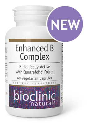


Learn More

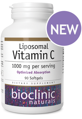
Learn More
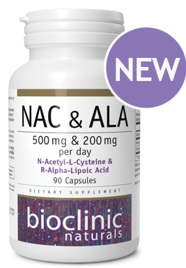
Learn More
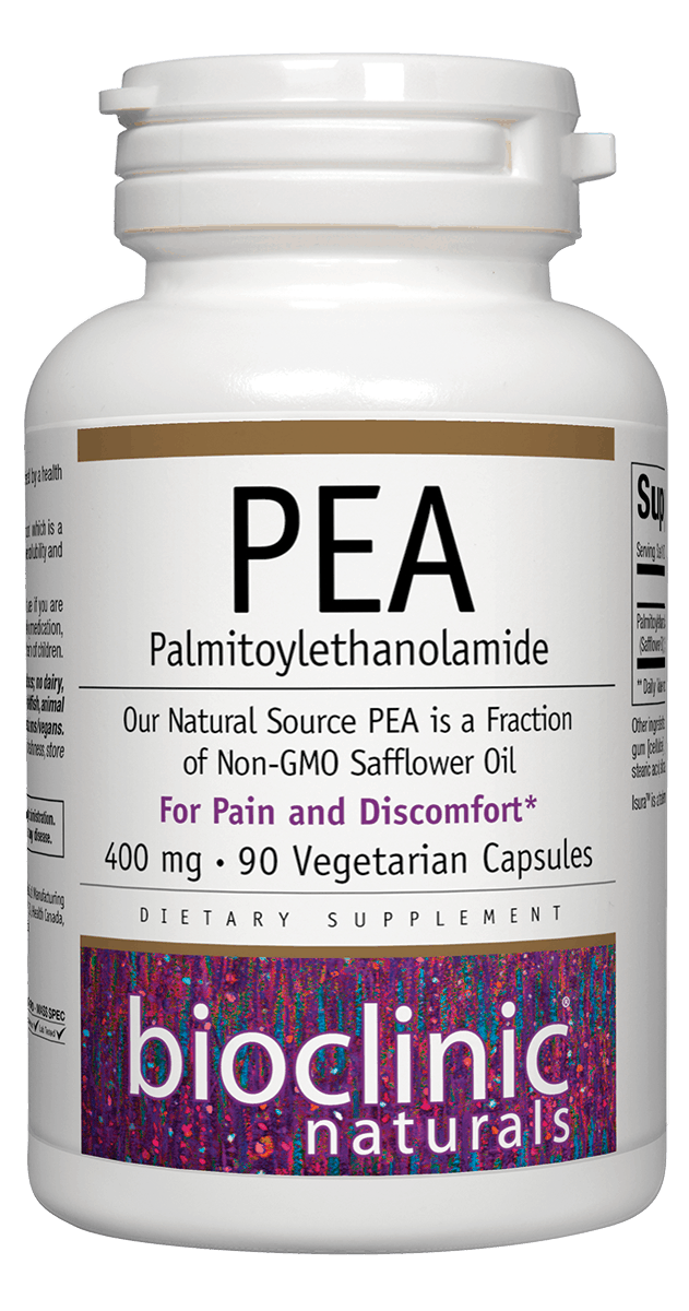
Find out More
Bioclinic Naturals products are ISURA certified to demonstrate our commitment to testing raw materials for quality, purity and potency.
At Bioclinic Naturals, we offer an exclusive health care practitioner line of
evidence-based natural health products.

WHAT WE OFFER
Every great product starts with the best ingredients. Our goal is to provide the best quality, tested raw materials, produced in state-of-the-art manufacturing facilities, and manufactured to the highest standards of quality in the world.

EXCLUSIVELY AVAILABLE TO HEALTH CARE PRACTITIONERS
Bioclinic Naturals provides products and educational tools for both practitioners and patients to support condition-specific challenges and overall health enhancement.
ABOUT BIOCLINIC NATURALS
We are Canada’s leading nutraceutical manufacturer. Our commitment to quality is reflected in our investments in expanding our in-house Quality Control, Quality Assurance, and Research & Development departments, and we have over 1000 acres of certified organic farmland in Canada’s Okanagan Valley.